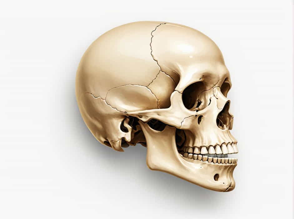
The zygomatic arch, commonly known as the cheekbone, is a key structural component of the human skull. It plays a crucial role in facial aesthetics, muscle attachment, and jaw movement. This bony arch is formed by the union of two bones:
- The zygomatic bone – Also called the cheekbone, this bone contributes to the prominence of the face.
- The temporal bone – Located at the sides of the skull, it houses structures important for hearing and balance.
Together, these bones create a strong yet lightweight structure that supports facial muscles and protects underlying tissues.
Anatomy of the Zygomatic Arch
The zygomatic arch is a curved bridge of bone that extends from the cheekbone to the side of the skull. It is primarily composed of two bony projections that meet and fuse:
1. The Temporal Process of the Zygomatic Bone
- A forward-extending projection from the zygomatic bone.
- Connects with the zygomatic process of the temporal bone.
2. The Zygomatic Process of the Temporal Bone
- A backward-extending projection from the temporal bone.
- Joins with the temporal process of the zygomatic bone to form the full arch.
Functions of the Zygomatic Arch
1. Provides Facial Structure and Contour
The zygomatic arch defines the cheekbones, contributing to the overall shape of the face. Well-developed zygomatic bones give the face a strong, prominent appearance, which is often associated with beauty and symmetry.
2. Supports the Muscles of Mastication (Chewing)
Several important muscles attach to the zygomatic arch, including:
- Masseter muscle – One of the strongest muscles involved in chewing.
- Temporalis muscle – Assists in jaw elevation and retraction.
These muscles work together to enable biting, chewing, and jaw movement.
3. Protects Facial Structures
The zygomatic arch acts as a protective shield, safeguarding important anatomical structures such as:
- The eyes and orbits (eye sockets).
- The temporal fossa, a region that houses muscles involved in jaw movement.
- The maxillary sinus, an air-filled space important for respiration.
4. Provides an Attachment Site for Ligaments
The zygomatic arch serves as an anchor point for several ligaments that help maintain jaw stability and facial support.
Common Injuries and Disorders of the Zygomatic Arch
1. Zygomatic Arch Fractures
- Often caused by sports injuries, falls, or car accidents.
- Symptoms include swelling, bruising, and difficulty opening the mouth.
- Severe fractures may require surgical realignment (open reduction and internal fixation).
2. Zygomaticomaxillary Complex (ZMC) Fractures
- A more severe type of injury involving multiple fractures in the zygomatic bone, maxilla, and orbital region.
- Can lead to facial asymmetry, vision problems, and nerve damage.
3. Temporomandibular Joint (TMJ) Dysfunction
- TMJ disorders can sometimes be linked to abnormal stress on the zygomatic arch.
- Symptoms include jaw pain, clicking sounds, and difficulty chewing.
How to Keep the Zygomatic Arch Healthy
1. Protect Against Facial Injuries
- Wear protective gear when engaging in sports or physical activities.
- Use seatbelts to prevent facial trauma in car accidents.
2. Strengthen Jaw and Facial Muscles
- Chewing exercises can help maintain muscle tone and function.
- Jaw stretches reduce tension and improve flexibility.
3. Maintain Good Posture and Jaw Alignment
- Avoid excessive jaw clenching or teeth grinding.
- Practice relaxation techniques to reduce facial muscle tension.
The zygomatic arch is formed by the zygomatic bone and the temporal bone, creating a strong and functional structure that supports facial shape, muscle attachment, and jaw movement. Keeping the zygomatic arch healthy is essential for maintaining facial aesthetics, chewing function, and overall skull stability. Understanding its anatomy and role can help prevent injuries and promote better facial health.