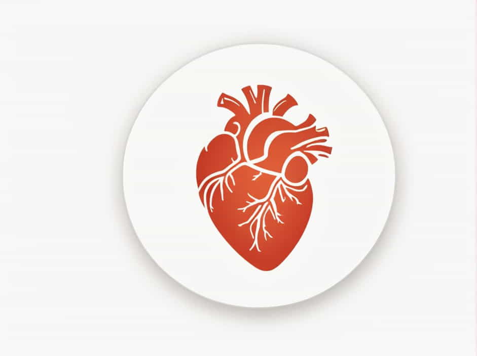
The aortic valve is a crucial component of the human heart, responsible for regulating blood flow between the left ventricle and the aorta. This valve ensures that oxygen-rich blood is efficiently pumped from the heart to the rest of the body.
Proper functioning of the aortic valve is essential for healthy circulation, preventing backflow of blood, and maintaining optimal heart performance. In this topic, we will explore the structure, function, common diseases, symptoms of dysfunction, and treatment options related to the aortic valve.
Anatomy of the Aortic Valve
Where Is the Aortic Valve Located?
The aortic valve is positioned at the junction of the left ventricle and the aorta, serving as a one-way gateway that controls blood movement from the heart’s main pumping chamber into the body’s largest artery.
Structure of the Aortic Valve
The aortic valve consists of:
- Three Cusps (Leaflets) – These thin, flexible flaps open and close to regulate blood flow.
- Annulus – A fibrous ring that provides structural support.
- Commissures – Points where the cusps attach to the valve wall.
While most people have a tricuspid aortic valve (three cusps), some individuals are born with a bicuspid aortic valve (two cusps), which may increase the risk of valve-related conditions.
Function of the Aortic Valve
The aortic valve plays a vital role in circulation by:
- Opening to Allow Blood Flow – During ventricular contraction (systole), the valve opens, allowing oxygen-rich blood to pass into the aorta.
- Closing to Prevent Backflow – When the heart relaxes (diastole), the valve closes, preventing blood from leaking back into the left ventricle.
This one-way mechanism ensures that the body receives a continuous supply of oxygenated blood without strain on the heart.
Common Aortic Valve Diseases
Despite its importance, the aortic valve can suffer from various disorders that affect heart function.
1. Aortic Stenosis
Aortic stenosis occurs when the valve becomes narrowed or stiff, restricting blood flow from the heart to the body. Causes include:
- Aging-related calcification (calcium buildup on the valve).
- Congenital defects (such as a bicuspid aortic valve).
- Rheumatic fever (causing scarring and thickening).
2. Aortic Regurgitation (Aortic Insufficiency)
Aortic regurgitation happens when the valve does not close properly, allowing blood to leak back into the left ventricle. This condition can lead to:
- Heart enlargement due to increased workload.
- Reduced blood supply to the body.
- Fatigue and breathlessness.
3. Congenital Aortic Valve Abnormalities
Some individuals are born with a malformed aortic valve, such as:
- Bicuspid Aortic Valve – A two-cusp valve instead of three.
- Unicuspid or Quadricuspid Valve – Less common variations that may cause complications later in life.
4. Endocarditis (Valve Infection)
Bacterial or fungal infections can damage the aortic valve, leading to life-threatening complications if untreated.
Symptoms of Aortic Valve Disease
Aortic valve dysfunction can develop gradually, with symptoms appearing as the condition worsens. Common signs include:
- Shortness of breath (especially during exertion).
- Chest pain or tightness.
- Fatigue and dizziness.
- Heart palpitations (irregular heartbeat).
- Swelling in the legs and ankles (due to fluid retention).
Severe cases may lead to heart failure, requiring immediate medical intervention.
Diagnosis of Aortic Valve Conditions
If aortic valve disease is suspected, doctors may perform several diagnostic tests, including:
1. Physical Examination
A doctor may listen for heart murmurs, an unusual sound caused by turbulent blood flow.
2. Echocardiogram
This ultrasound test visualizes the heart’s structure and function, detecting valve abnormalities.
3. Electrocardiogram (ECG/EKG)
An ECG records the heart’s electrical activity, identifying irregular rhythms.
4. Cardiac MRI or CT Scan
Advanced imaging techniques provide detailed heart anatomy to assess valve damage.
5. Cardiac Catheterization
A small catheter is inserted into a blood vessel to measure heart pressure and function.
Treatment Options for Aortic Valve Disease
Treatment depends on the severity of the condition and may include medications, lifestyle changes, or surgery.
1. Medications
While medications cannot cure valve disease, they help manage symptoms and reduce complications. Common drugs include:
- Beta-blockers (to control heart rate and blood pressure).
- Diuretics (to reduce fluid buildup).
- Blood thinners (to prevent clots in patients with valve replacement).
2. Lifestyle Modifications
- Heart-healthy diet (low in saturated fat and sodium).
- Regular exercise (as recommended by a doctor).
- Avoid smoking and excessive alcohol consumption.
3. Surgical Treatments
Severe aortic valve disease often requires surgical intervention.
Aortic Valve Replacement (AVR)
A damaged valve is replaced with a prosthetic valve, which can be:
- Mechanical valve (made of metal and plastic, requires lifelong blood thinners).
- Biological valve (made from pig, cow, or human tissue, but may wear out over time).
Transcatheter Aortic Valve Replacement (TAVR)
A minimally invasive alternative to open-heart surgery, TAVR is often recommended for high-risk patients.
Preventing Aortic Valve Disease
While some causes of valve disease are genetic or age-related, certain steps can help maintain aortic valve health:
- Control Blood Pressure and Cholesterol – Reducing strain on the heart helps prevent valve thickening and hardening.
- Treat Strep Infections Promptly – Preventing rheumatic fever reduces the risk of future valve complications.
- Regular Heart Check-Ups – Early detection of heart murmurs or irregularities allows for better management.
- Maintain a Heart-Healthy Lifestyle – Balanced nutrition, exercise, and stress management contribute to overall cardiovascular health.
Interesting Facts About the Aortic Valve
- The aortic valve opens and closes approximately 100,000 times per day.
- It withstands high pressure as it pumps blood into the aorta at great force.
- Some professional athletes are diagnosed with bicuspid aortic valve and continue competing with proper monitoring.
- The first successful aortic valve replacement surgery was performed in the 1960s, revolutionizing heart treatment.
The aortic valve is a critical component of the cardiovascular system, ensuring efficient blood flow from the left ventricle to the aorta. Understanding its function, recognizing early signs of disease, and seeking timely treatment are essential for heart health and overall well-being.
With advancements in medical technology, diagnosis, and treatment options, individuals with aortic valve disorders can lead healthy, active lives when properly managed. Regular heart screenings, a balanced lifestyle, and early medical intervention play key roles in preventing complications and maintaining optimal cardiovascular function.
