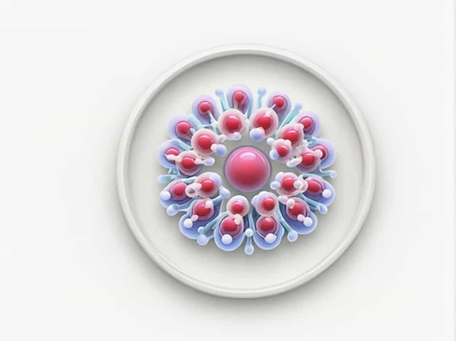
Cells are the fundamental units of life, and within them are specialized structures called organelles that perform essential functions. Understanding the ultrastructure of cell organelles is crucial for studying cell biology, disease mechanisms, and molecular functions. But how can we study these tiny structures in detail?
This topic explores the best techniques to study the ultrastructure of cell organelles, including electron microscopy, X-ray crystallography, and fluorescence microscopy.
What Is the Ultrastructure of Cell Organelles?
Definition of Ultrastructure
The ultrastructure of a cell refers to the fine details of its components, visible only at very high magnification. This includes the membranes, internal structures, and molecular compositions of organelles like the nucleus, mitochondria, Golgi apparatus, endoplasmic reticulum, and lysosomes.
Why Is Studying the Ultrastructure Important?
- Helps understand cellular functions at a molecular level.
- Identifies disease-related abnormalities in organelles.
- Aids in drug development and genetic research.
- Provides insight into evolutionary biology.
Techniques Used to Study the Ultrastructure of Cell Organelles
1. Electron Microscopy (EM)
One of the most powerful tools for studying cell ultrastructure is electron microscopy. Unlike light microscopes, electron microscopes use electron beams instead of light, allowing for much higher magnification and resolution.
Types of Electron Microscopy
A. Transmission Electron Microscopy (TEM)
- Provides detailed internal structure of organelles.
- Electrons pass through thin cell sections to create high-resolution images.
- Used to study the mitochondria, endoplasmic reticulum, and nuclear membrane.
B. Scanning Electron Microscopy (SEM)
- Produces 3D images of cell surfaces and organelles.
- Electrons bounce off the specimen to generate images.
- Useful for studying the outer structures of organelles.
2. X-ray Crystallography
- Used to analyze the molecular structure of proteins within organelles.
- Helps determine the shape and function of organelle components.
- Essential for studying ribosomes, enzymes, and membrane proteins.
3. Fluorescence Microscopy
- Uses fluorescent dyes that bind to specific organelles.
- Allows visualization of live cells without damaging them.
- Commonly used for studying the Golgi apparatus, lysosomes, and mitochondria.
4. Confocal Microscopy
- A type of fluorescence microscopy that provides sharper images.
- Uses a laser scanning system to focus on specific cell layers.
- Helpful for studying dynamic changes in organelles.
5. Cryo-Electron Microscopy (Cryo-EM)
- A revolutionary technique that preserves samples in frozen states.
- Allows observation of biological molecules in their natural environment.
- Used for studying ribosomes, viruses, and cellular membranes.
How These Techniques Help Study Specific Organelles
1. Studying the Nucleus
- TEM helps reveal the nuclear envelope, chromatin structure, and nucleolus.
- Fluorescence microscopy highlights DNA and RNA activity.
2. Analyzing Mitochondria
- TEM provides detailed images of the inner and outer mitochondrial membranes.
- Fluorescence microscopy tracks ATP production and mitochondrial function.
3. Observing the Golgi Apparatus
- Confocal microscopy helps visualize the Golgi’s role in protein modification.
- TEM captures detailed Golgi cisternae structures.
4. Investigating the Endoplasmic Reticulum (ER)
- TEM reveals the difference between smooth and rough ER.
- X-ray crystallography helps study the ribosome structure on rough ER.
5. Exploring Lysosomes and Peroxisomes
- Fluorescence microscopy tracks enzyme activity inside lysosomes.
- Cryo-EM provides molecular details of peroxisomal proteins.
Applications of Studying Cell Ultrastructure
1. Medical Research
- Helps in cancer detection by identifying changes in nuclear structure.
- Aids in diagnosing mitochondrial diseases.
2. Drug Development
- Identifies drug targets within organelles.
- Helps develop antibiotics that affect bacterial ribosomes.
3. Genetic Engineering
- Provides insight into DNA modifications.
- Supports research in gene therapy and CRISPR technology.
4. Biotechnology and Agriculture
- Helps improve crop genetics by studying plant cell ultrastructure.
- Supports the development of bioengineered food.
Studying the ultrastructure of cell organelles is essential for understanding cell biology, disease mechanisms, and biotechnology. Advanced techniques like electron microscopy, X-ray crystallography, and fluorescence microscopy have revolutionized how scientists explore these microscopic structures.
By continuing to refine these methods, researchers can uncover new insights into how cells function, how diseases develop, and how we can develop new medical treatments.
