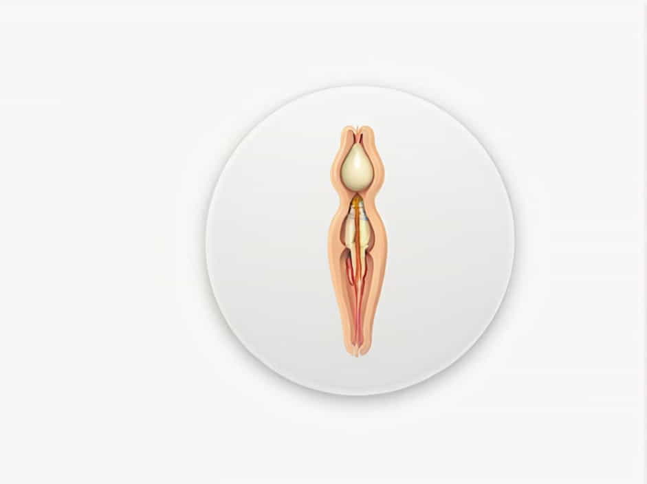
The incisive canal and the transverse palatine suture are two important anatomical structures of the hard palate. The incisive canal serves as a passage for nerves and blood vessels, while the transverse palatine suture marks the junction between the maxillary and palatine bones. Understanding the relationship between these structures is essential for dentists, surgeons, and anyone studying craniofacial anatomy.
Anatomy of the Incisive Canal
The incisive canal, also known as the nasopalatine canal, is a bony passage located in the anterior portion of the hard palate. It extends from the nasal cavity to the oral cavity, opening at the incisive foramen behind the central incisors.
Location and Structure
- The incisive canal is positioned within the premaxilla, just posterior to the upper central incisors.
- It consists of two bony tunnels that converge into a single opening at the incisive foramen.
- The canal houses the nasopalatine nerve and branches of the sphenopalatine artery, which supply sensation and blood flow to the anterior palate.
Functions of the Incisive Canal
- Allows the nasopalatine nerve to provide sensory innervation to the anterior hard palate.
- Transmits small blood vessels that contribute to vascular supply.
- Plays a role in maxillofacial surgeries and dental procedures, especially in implant placement.
Anatomy of the Transverse Palatine Suture
The transverse palatine suture is a bony junction located in the posterior part of the hard palate. It marks the articulation between the maxillary processes and the horizontal plates of the palatine bones.
Location and Structure
- The suture runs horizontally across the hard palate, separating the anterior maxilla from the posterior palatine bones.
- It forms part of the mid-palatal region, playing a crucial role in craniofacial stability.
- In some individuals, the transverse palatine suture may be slightly irregular, which can affect dental occlusion and palatal expansion procedures.
Functions of the Transverse Palatine Suture
- Provides structural integrity to the hard palate.
- Serves as a reference point for orthodontic and surgical procedures.
- Allows for growth and fusion of the palatal bones during development.
Relationship Between the Incisive Canal and the Transverse Palatine Suture
The incisive canal and transverse palatine suture are anatomically related but serve different functions.
1. Proximity in the Hard Palate
- The incisive canal is positioned in the anterior portion of the hard palate.
- The transverse palatine suture is located posteriorly, separating the maxilla from the palatine bones.
- Despite their different locations, both structures contribute to the stability and function of the hard palate.
2. Clinical Importance in Surgeries and Dental Procedures
- In dental implant procedures, avoiding the incisive canal is crucial to prevent damage to the nasopalatine nerve.
- Palatal expansion procedures may involve the transverse palatine suture, requiring careful consideration of bone fusion and flexibility.
3. Developmental and Anatomical Variations
- Some individuals may have variations in the size and shape of the incisive canal, affecting nerve pathways.
- The transverse palatine suture can exhibit asymmetry or irregular fusion, influencing orthodontic treatments.
Clinical Significance of the Incisive Canal and Transverse Palatine Suture
Understanding these structures is essential for various medical and dental fields.
1. Dental Implant Placement
- The incisive canal must be avoided to prevent nerve damage and postoperative complications.
- Careful radiographic evaluation is necessary before implant placement in the anterior maxilla.
2. Maxillofacial Surgeries
- Procedures like palatal expansion and cleft palate repair require an understanding of the transverse palatine suture.
- In cases of midface trauma, surgeons must assess the structural integrity of the palate to ensure proper healing.
3. Orthodontic Treatments
- Palatal expansion procedures often target the transverse palatine suture to widen the upper jaw.
- Orthodontists must consider the ossification and fusion state of this suture, especially in older patients.
Maintaining Oral and Palatal Health
To prevent complications related to the incisive canal and transverse palatine suture, maintaining good oral health is essential.
1. Regular Dental Check-Ups
- Early detection of bone irregularities can prevent future dental complications.
- Dentists assess the incisive canal and palatal sutures during routine exams.
2. Proper Oral Hygiene
- Brushing and flossing prevent gingival inflammation, which can affect palatal health.
- Using fluoride-based toothpaste helps strengthen bone structures.
3. Orthodontic and Surgical Consultations
- If palatal expansion or dental implants are needed, consulting a specialist ensures successful outcomes.
- 3D imaging techniques allow precise evaluation of bone structure and nerve positioning.
The incisive canal and transverse palatine suture are fundamental structures in craniofacial anatomy. While the incisive canal serves as a passageway for nerves and blood vessels, the transverse palatine suture provides structural support to the hard palate. Their relationship is essential in dental, orthodontic, and maxillofacial procedures, making their study crucial for medical professionals. Proper understanding of these structures enhances clinical outcomes, ensuring both functional and aesthetic success in various treatments.
