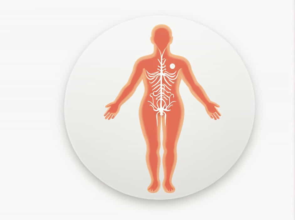
The pelvic viscera include important organs such as the bladder, rectum, uterus, prostate, and reproductive structures. These organs receive autonomic and somatic nerve supply, which controls their function, including urination, defecation, and sexual responses.
Understanding the nerve supply of the pelvic viscera is crucial for diagnosing and managing conditions like pelvic pain, incontinence, and nerve damage. This topic provides a detailed overview of the nerves involved and their clinical significance.
Overview of Pelvic Nerve Supply
The nerve supply of the pelvic viscera comes from two main systems:
-
Autonomic Nervous System (ANS)
- Sympathetic nerves (T10-L2) – Control blood flow and inhibit bladder contraction.
- Parasympathetic nerves (S2-S4) – Promote bladder emptying and rectal contraction.
-
Somatic Nervous System
- Provides voluntary control over structures like the external anal and urethral sphincters.
Autonomic Nervous System of the Pelvic Viscera
1. Sympathetic Nerve Supply
The sympathetic nervous system arises from the thoracolumbar region (T10-L2) and reaches the pelvic organs through the hypogastric plexuses.
Functions of Sympathetic Nerves
- Inhibit bladder contraction (preventing urination).
- Promote internal urethral and anal sphincter contraction.
- Control ejaculation in males.
- Regulate blood flow to pelvic organs.
Pathway of Sympathetic Nerves
- Sympathetic fibers travel from T10-L2 to the superior hypogastric plexus.
- They descend to the inferior hypogastric plexus, where they innervate pelvic organs.
2. Parasympathetic Nerve Supply
The parasympathetic nervous system originates from the sacral spinal cord (S2-S4) and travels through the pelvic splanchnic nerves.
Functions of Parasympathetic Nerves
- Stimulate bladder contraction (promoting urination).
- Relax the internal urethral sphincter.
- Stimulate peristalsis in the rectum (promoting defecation).
- Control erectile function in males and females.
Pathway of Parasympathetic Nerves
- Parasympathetic fibers travel from S2-S4 through the pelvic splanchnic nerves.
- They reach the inferior hypogastric plexus and supply the bladder, rectum, and reproductive organs.
Somatic Nerve Supply of the Pelvic Viscera
The somatic nervous system provides voluntary control over some pelvic structures, primarily through the pudendal nerve (S2-S4).
Pudendal Nerve
- Originates from S2-S4.
- Provides motor control to the external anal sphincter and external urethral sphincter.
- Carries sensory information from the perineum, penis, and clitoris.
Functions of the Pudendal Nerve
- Allows voluntary control of urination and defecation.
- Contributes to sexual sensation and function.
- Important for pelvic floor muscle strength.
Pelvic Nerve Plexuses and Their Role
1. Superior Hypogastric Plexus
- Located near the aortic bifurcation.
- Contains sympathetic fibers from T10-L2.
- Sends branches to the inferior hypogastric plexus.
2. Inferior Hypogastric Plexus
- Located on both sides of the pelvis.
- Contains both sympathetic and parasympathetic fibers.
- Supplies the bladder, rectum, prostate, uterus, and vagina.
3. Pelvic Splanchnic Nerves
- Carry parasympathetic fibers from S2-S4.
- Promote bladder contraction and rectal motility.
4. Sacral Plexus (L4-S4)
- Gives rise to the pudendal nerve, which controls the perineum and pelvic floor muscles.
Clinical Significance of Pelvic Nerve Supply
1. Bladder Dysfunction
- Overactive bladder: Due to excessive parasympathetic activity.
- Urinary retention: Due to damage to parasympathetic nerves (S2-S4).
2. Fecal Incontinence
- Weakness of the pudendal nerve can impair voluntary control over the anal sphincter.
3. Erectile Dysfunction
- Damage to the parasympathetic nerves (S2-S4) affects erection.
- Sympathetic dysfunction can impair ejaculation.
4. Chronic Pelvic Pain
- Irritation of the pudendal nerve can cause pudendal neuralgia, leading to perineal pain.
5. Childbirth-Related Nerve Damage
- Prolonged labor can compress the pudendal nerve, leading to temporary incontinence or pelvic floor weakness.
The nerve supply of the pelvic viscera involves a complex interaction between sympathetic, parasympathetic, and somatic nerves. These nerves regulate essential functions such as urination, defecation, and sexual activity.
Understanding these nerve pathways is crucial for diagnosing and treating pelvic disorders, nerve injuries, and chronic pain conditions.
