The meningeal branch of the mandibular nerve is a small but essential nerve that plays a crucial role in the nervous system. It is responsible for innervating the dura mater in the cranial cavity and providing sensory function. This branch, also known as the nervus spinosus, originates from the mandibular nerve (V3), which is the largest division of the trigeminal nerve (cranial nerve V).
Understanding the anatomy, function, and clinical relevance of this nerve is essential for medical professionals, especially those specializing in neurology, dentistry, and maxillofacial surgery. This topic will provide a detailed exploration of the meningeal branch of the mandibular nerve, including its course, role, and associated medical conditions.
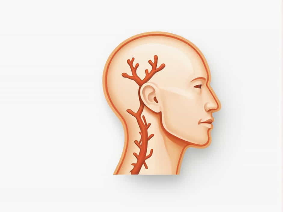
Anatomy of the Meningeal Branch of the Mandibular Nerve
Origin and Course
The meningeal branch (nervus spinosus) originates from the mandibular nerve (V3), which emerges from the foramen ovale in the skull. Shortly after its exit, this nerve re-enters the cranial cavity through the foramen spinosum alongside the middle meningeal artery.
Once inside the cranial cavity, the nerve branches out and supplies the dura mater of the middle cranial fossa, contributing to the sensory innervation of the meninges.
Relation to Other Structures
- The mandibular nerve (V3) is a mixed nerve containing both motor and sensory fibers.
- The meningeal branch is purely sensory, meaning it does not control any muscles but instead transmits sensory information.
- The nerve travels with the middle meningeal artery, making this anatomical relationship significant in head trauma cases involving vascular injuries.
Functions of the Meningeal Branch
The primary function of the meningeal branch is to provide sensory innervation to the dura mater in the middle cranial fossa. This helps in:
- Pain perception: The dura mater contains pain receptors, and the meningeal branch plays a role in headache sensations caused by meningeal irritation.
- Protective function: The dura mater serves as a protective covering for the brain, and its innervation is crucial for detecting injuries or pressure changes.
- Neural communication: It works in coordination with other branches of the trigeminal nerve to transmit sensory input from the skull and brain coverings.
Clinical Significance of the Meningeal Branch of the Mandibular Nerve
1. Headaches and Neuralgia
Since the meningeal branch is responsible for dura mater sensation, irritation or compression of this nerve can lead to headaches or neuralgic pain.
- Tension headaches and migraines may involve this nerve due to meningeal inflammation.
- Trigeminal neuralgia, a condition affecting the trigeminal nerve, can sometimes involve the meningeal branch, leading to sharp, stabbing pain in the head.
2. Middle Meningeal Artery Rupture and Epidural Hematoma
Due to its close association with the middle meningeal artery, any injury to this artery, such as in a skull fracture, can affect the meningeal branch as well.
- Epidural hematoma is a severe condition that results from middle meningeal artery rupture.
- Symptoms include severe headaches, loss of consciousness, and neurological deficits.
- Damage to the meningeal branch in such cases can contribute to sensory abnormalities in the cranial dura mater.
3. Compression Due to Tumors or Lesions
- Tumors in the middle cranial fossa may compress the meningeal branch, leading to persistent headaches.
- Conditions such as meningiomas or trigeminal schwannomas can put pressure on this nerve, affecting its function.
4. Mandibular Nerve Blocks in Dentistry
The mandibular nerve (V3) is often targeted in dental anesthesia. While the meningeal branch is not directly involved in dental procedures, understanding its pathway is crucial to avoiding complications during nerve blocks.
Comparison with Other Meningeal Nerve Branches
The trigeminal nerve (CN V) has three divisions, and each provides meningeal branches:
| Nerve Division | Meningeal Branch | Innervated Area |
|---|---|---|
| Ophthalmic (V1) | Tentorial nerve | Dura of anterior cranial fossa |
| Maxillary (V2) | Middle meningeal branch | Dura of middle cranial fossa |
| Mandibular (V3) | Nervus spinosus | Dura of middle cranial fossa |
Among these, the meningeal branch of the mandibular nerve is unique due to its re-entry into the cranial cavity through the foramen spinosum.
How to Maintain Nerve Health
While nerve disorders are not always preventable, some general health practices can help maintain nerve function and prevent complications:
1. Maintain Proper Posture
Poor posture, especially prolonged neck bending, can contribute to nerve compression, leading to headaches and discomfort.
2. Avoid Head Injuries
Wearing helmets during activities like cycling, motorcycling, or contact sports can protect the skull and reduce the risk of trauma affecting the middle meningeal artery and nerve.
3. Stay Hydrated and Eat a Balanced Diet
- B vitamins (B1, B6, B12) are crucial for nerve function.
- Drinking enough water helps maintain circulation, preventing dehydration-related headaches.
4. Manage Stress
- Chronic stress can increase muscle tension, potentially affecting nerve function.
- Relaxation techniques such as meditation, yoga, and deep breathing can help prevent tension-related headaches.
5. Seek Medical Attention for Persistent Headaches
If you experience chronic headaches, unexplained pain, or sensory disturbances, consult a neurologist or healthcare professional to rule out underlying nerve involvement.
The meningeal branch of the mandibular nerve is a small but vital sensory nerve that plays an important role in the innervation of the dura mater in the middle cranial fossa. Despite its size, it has significant clinical relevance, especially in conditions related to headaches, nerve compression, and traumatic brain injuries.
By understanding its anatomy, functions, and clinical significance, medical professionals and students can appreciate its role in neurology and craniofacial anatomy. Proper care, including injury prevention, stress management, and medical attention for persistent symptoms, can help maintain optimal nerve health.