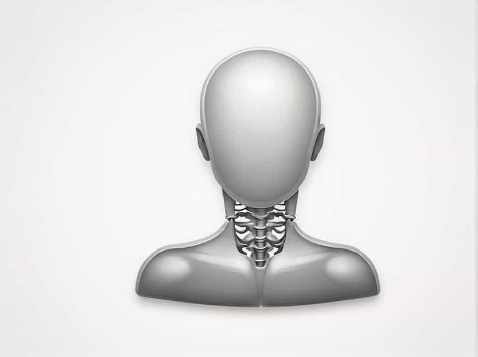
The uppermost vertebrae in the neck play a crucial role in supporting the skull, enabling head movement, and protecting the spinal cord. These vertebrae are part of the cervical spine, which consists of seven vertebrae labeled C1 to C7. The first two cervical vertebrae, known as the atlas (C1) and axis (C2), are the most important for head movement and stability.
Understanding the anatomy and function of these vertebrae is essential for learning about neck mobility, posture, and potential spinal injuries. This topic explores the structure, role, and clinical significance of the uppermost cervical vertebrae.
The Cervical Spine: An Overview
The cervical spine is the uppermost section of the vertebral column and consists of seven vertebrae (C1-C7). It connects the skull to the thoracic spine and allows for a wide range of head movements.
Key Functions of the Cervical Spine
- Supports the weight of the head (which weighs around 4.5 to 5 kg).
- Allows movement of the head in multiple directions.
- Protects the spinal cord as it exits the brainstem.
- Facilitates blood flow to the brain through the vertebral arteries.
The uppermost vertebrae—C1 (Atlas) and C2 (Axis)—are structurally different from the other cervical vertebrae and have unique roles in neck mobility.
C1 (Atlas): The First Cervical Vertebra
Structure of the Atlas
The atlas (C1) is the first cervical vertebra and is named after Atlas from Greek mythology, who carried the heavens on his shoulders. Unlike other vertebrae, the atlas lacks a vertebral body and spinous process. Instead, it has:
- Anterior and posterior arches – Forming a ring-like structure.
- Lateral masses – Thickened areas that articulate with the skull.
- Superior articular facets – Where the occipital condyles of the skull rest, forming the atlanto-occipital joint.
Function of the Atlas
The atlas vertebra is responsible for:
- Supporting the skull and connecting it to the spine.
- Facilitating the nodding motion (“yes” movement) of the head.
- Providing a passage for the spinal cord and vertebral arteries.
Clinical Relevance: Atlas-Related Conditions
- Atlantoaxial Instability – A condition where excessive movement occurs between C1 and C2, often seen in people with Down syndrome or rheumatoid arthritis.
- Jefferson Fracture – A break in the anterior or posterior arch of C1, usually due to a severe impact to the head.
C2 (Axis): The Second Cervical Vertebra
Structure of the Axis
The axis (C2) is the second cervical vertebra and has a unique odontoid process (dens), which extends upward into the atlas. This structure allows for rotational movement of the head.
Function of the Axis
- Enables head rotation (“no” movement) through the atlantoaxial joint.
- Provides a pivot point for the atlas to rotate around.
- Supports the skull while maintaining spinal stability.
Clinical Relevance: Axis-Related Conditions
- Hangman’s Fracture – A fracture of C2, often caused by hyperextension of the neck.
- Odontoid Fracture – A break in the dens, which can be life-threatening if it compresses the spinal cord.
Atlanto-Occipital and Atlantoaxial Joints
The uppermost vertebrae in the neck function through two key joints:
- Atlanto-Occipital Joint (C0-C1) – Connects the skull (occipital bone) to the atlas (C1), allowing nodding motions.
- Atlantoaxial Joint (C1-C2) – Connects the atlas (C1) to the axis (C2), enabling head rotation.
These joints provide stability and flexibility, making neck movement possible.
C3 to C7: The Lower Cervical Vertebrae
While C1 and C2 are the most specialized vertebrae in the cervical spine, C3 to C7 also contribute to neck movement and spinal protection.
Features of C3 to C7 Vertebrae
- Smaller bodies compared to thoracic and lumbar vertebrae.
- Bifid spinous processes (except for C7, which has a prominent spinous process).
- Foramina in transverse processes that allow vertebral arteries to pass through.
Functions of the Lower Cervical Vertebrae
- Allow flexion, extension, and lateral bending of the neck.
- Protect the spinal cord and nerve roots.
- Serve as attachment points for muscles and ligaments.
Clinical Relevance: Common Cervical Spine Disorders
- Cervical Spondylosis – Age-related degeneration of the cervical spine, leading to pain and stiffness.
- Herniated Cervical Disc – A slipped or bulging disc that can press on nerves, causing pain or numbness.
- Whiplash Injury – Sudden hyperextension and hyperflexion of the neck, often due to car accidents.
Importance of the Upper Cervical Vertebrae
The atlas (C1) and axis (C2) are among the most important vertebrae in the human body. They allow for head movement, protect the brainstem, and contribute to overall spinal stability.
Why Are These Vertebrae Unique?
- Atlas (C1) has no vertebral body, making it lightweight yet strong.
- Axis (C2) has the odontoid process (dens), which acts as a pivot point for head rotation.
- They form joints that allow nearly 50% of head movement.
Injuries or abnormalities in these vertebrae can lead to serious neurological issues, including paralysis or difficulty breathing.
The uppermost vertebrae in the neck, C1 (Atlas) and C2 (Axis), play a vital role in head movement, spinal protection, and neurological function.
- The atlas (C1) supports the skull and enables nodding motions.
- The axis (C2) allows for head rotation, providing stability and flexibility.
- Together, they form crucial joints that allow a wide range of motion.
Understanding the structure and function of these vertebrae helps in recognizing spinal health issues, preventing injuries, and improving posture. Proper care of the cervical spine is essential for maintaining overall spinal health and mobility.
