Acetabular Fractures: What Radiologists Should Know
Acetabular fractures, which involve the socket of the hip joint, are complex injuries often resulting from high-energy trauma. These fractures can significantly impact a patient’s mobility and quality of life if not properly diagnosed and treated. Radiologists play a critical role in the assessment and management of acetabular fractures. This article will provide a comprehensive overview of what radiologists should know about acetabular fractures, including their classification, imaging techniques, and key considerations in diagnosis.
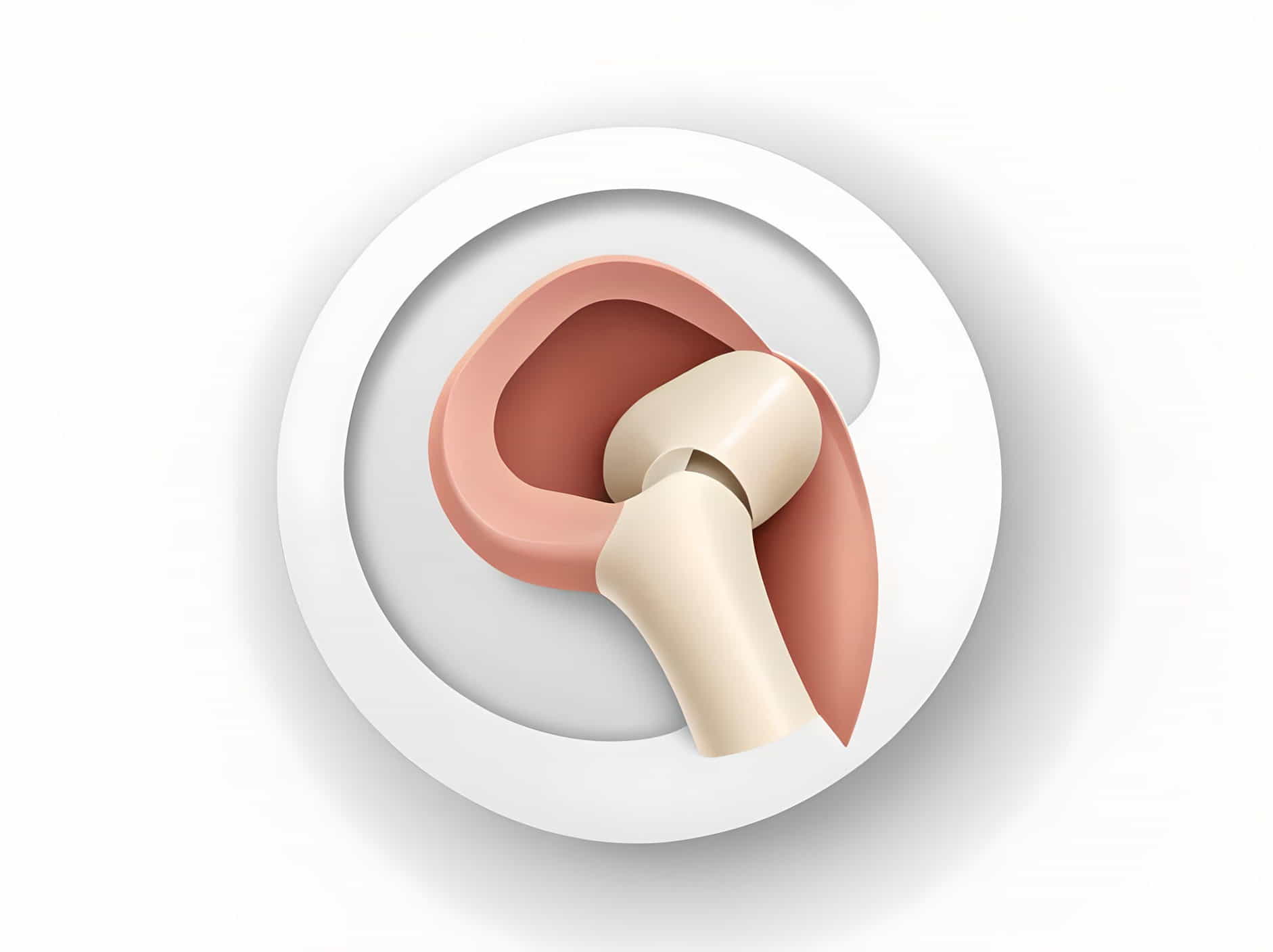
Anatomy of the Acetabulum
Understanding the anatomy of the acetabulum is crucial for accurately diagnosing and evaluating fractures. The acetabulum is a deep, cup-shaped structure that forms part of the hip joint. It is composed of three bones:
- Ilium: The uppermost and largest part of the pelvis.
- Ischium: The lower and back part of the pelvis.
- Pubis: The front part of the pelvis.
These bones converge to form the acetabular fossa, which articulates with the femoral head, creating the hip joint.
Classification of Acetabular Fractures
Acetabular fractures are classified based on the involvement of specific anatomical regions and fracture patterns. The most widely used classification system is the Letournel-Judet classification, which divides fractures into two main categories: elementary and associated.
Elementary Fractures
- Posterior Wall: The most common type, involving the back wall of the acetabulum.
- Posterior Column: Fractures involving the entire back column.
- Anterior Wall: Fractures of the front wall.
- Anterior Column: Fractures involving the front column.
- Transverse: Fractures that run horizontally through the acetabulum.
Associated Fractures
- Both Columns: Fractures involving both the anterior and posterior columns.
- T-Shaped: A transverse fracture combined with a vertical component.
- Anterior with Posterior Hemi-transverse: Involving the anterior column with a transverse extension.
- Posterior with Anterior Hemi-transverse: Involving the posterior column with a transverse extension.
Imaging Techniques
Accurate imaging is essential for diagnosing acetabular fractures and planning appropriate treatment. Radiologists should be familiar with various imaging modalities and their applications.
Plain Radiography
Plain radiographs are typically the first imaging modality used. Key views include:
- Anteroposterior (AP) View: Provides an overview of the pelvis and hip joint.
- Judet Views: Oblique views (iliac and obturator oblique) that help visualize the anterior and posterior columns of the acetabulum.
Computed Tomography (CT)
CT scans are the gold standard for detailed evaluation of acetabular fractures. They provide three-dimensional reconstructions that allow for precise assessment of fracture patterns, displacement, and the involvement of articular surfaces.
- Axial, Coronal, and Sagittal Views: These views help in understanding the extent and complexity of the fracture.
- 3D Reconstruction: Offers a comprehensive view of the fracture, aiding in surgical planning.
Magnetic Resonance Imaging (MRI)
MRI is not routinely used for diagnosing acetabular fractures but can be valuable in specific cases, such as:
- Assessing Soft Tissue Injuries: MRI can evaluate associated soft tissue injuries, including labral tears and cartilage damage.
- Occult Fractures: MRI can detect fractures not visible on plain radiographs or CT scans.
Key Considerations in Diagnosis
Radiologists should be aware of several key considerations when diagnosing acetabular fractures:
Fracture Pattern Recognition
Understanding the fracture pattern is crucial for accurate classification and treatment planning. This involves recognizing specific fracture lines and their involvement with the acetabular columns and walls.
Displacement and Comminution
Evaluating the degree of displacement and comminution (fragmentation) of the fracture is essential. Significant displacement can affect joint congruence and stability, influencing the treatment approach.
Articular Surface Involvement
Assessing the involvement of the articular surface is critical, as this impacts the risk of post-traumatic arthritis. Even small fragments within the joint can lead to significant long-term issues if not addressed.
Associated Injuries
High-energy trauma causing acetabular fractures often results in associated injuries. Radiologists should be vigilant for:
- Pelvic Ring Fractures: These can accompany acetabular fractures and complicate management.
- Vascular and Nerve Injuries: These are less common but can have serious implications for the patient’s recovery.
Acetabular fractures are complex injuries that require a thorough understanding of anatomy, classification, and imaging techniques for accurate diagnosis and effective management. Radiologists play a pivotal role in identifying these fractures and guiding treatment plans. By recognizing fracture patterns, assessing displacement and articular involvement, and being aware of associated injuries, radiologists can significantly impact patient outcomes. Mastery of imaging modalities, particularly CT, is essential for detailed evaluation and successful management of acetabular fractures.